A teoria da origem humana na África é frequentemente questionada: bom para a Ciência!

segunda-feira, outubro 30, 2017

The theory that humans emerged in Africa is often questioned. That’s good for science

 
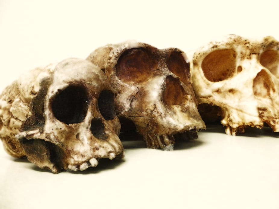
The Taung child (foreground) was the first of a long series of human ancestors discovered in Africa. Julien Benoit
 
 For nearly a century now the African root of human evolutionary theory has remained strong and unbowed. It is proved by a tremendous fossil record that documents the diversity of hominoids – apes and their relatives – across the continent through tens of millions of years. 

Then, the human branch of the evolutionary tree (hominins) split only seven or eight million years ago from our closest ape relatives, chimpanzees and gorillas. The oldest recorded hominin, whose skull was found in Chad and has been nicknamed Toumaï, is just a little younger than this. 

Africa remained the unique centre of hominin evolution for approximately the next six million years. 1.8 million years ago, Homo erectus first left the continent – and today we’re everywhere.

Several recent pieces of research have questioned this established consensus. They have, either directly or in media articles about the work, suggested that humankind’s evolutionary tree should be re-rooted in Europe. This is the nature of science: a paradigm that cannot be questioned on a regular basis becomes a dogma. 

So let’s examine these so-called “paradigm shifters” and see whether Africa should be stripped of the title of “cradle of humankind”.
...

Read more here/Leia mais aqui: The Conversation

O Atlas das Células Humanas: Abordagens e desafios técnicos

domingo, outubro 29, 2017

The Human Cell Atlas: Technical approaches and challenges

Chung-Chau Hon Jay W Shin Piero Carninci Michael JT Stubbington

Briefings in Functional Genomics, elx029, https://doi.org/10.1093/bfgp/elx029

Published: 28 October 2017


Abstract

The Human Cell Atlas is a large, international consortium that aims to identify and describe every cell type in the human body. The comprehensive cellular maps that arise from this ambitious effort have the potential to transform many aspects of fundamental biology and clinical practice. Here, we discuss the technical approaches that could be used today to generate such a resource and also the technical challenges that will be encountered.

Human Cell Atlas, single cell, RNA sequencing, bioinformatics

Darwin, mais complexidade: um modelo estrutural de mudança de filamentos flagelares em múltiplas espécies de bactérias

sexta-feira, outubro 27, 2017

A structural model of flagellar filament switching across multiple bacterial species

Fengbin Wang, Andrew M. Burrage, Sandra Postel, Reece E. Clark, Albina Orlova, Eric J. Sundberg, Daniel B. Kearns & Edward H. Egelman

Nature Communications 8, Article number: 960 (2017)


Download Citation

Bacterial structural biologyCellular motility Cryoelectron microscopy Supramolecular assembly

Received: 05 July 2017 Accepted: 15 August 2017 Published online: 16 October 2017


Abstract

The bacterial flagellar filament has long been studied to understand how a polymer composed of a single protein can switch between different supercoiled states with high cooperativity. Here we present near-atomic resolution cryo-EM structures for flagellar filaments from both Gram-positive Bacillus subtilis and Gram-negative Pseudomonas aeruginosa. Seven mutant flagellar filaments in B. subtilis and two in P. aeruginosa capture two different states of the filament. These reliable atomic models of both states reveal conserved molecular interactions in the interior of the filament among B. subtilis, P. aeruginosa and Salmonella enterica. Using the detailed information about the molecular interactions in two filament states, we successfully predict point mutations that shift the equilibrium between those two states. Further, we observe the dimerization of P. aeruginosa outer domains without any perturbation of the conserved interior of the filament. Our results give new insights into how the flagellin sequence has been “tuned” over evolution.

Acknowledgements

This work was supported by NIH GM122510 (to E.H.E.) and GM093030 (to D.B.K.). The cryo-EM work was conducted at the Molecular Electron Microscopy Core facility at the University of Virginia, which is supported by the School of Medicine and built with NIH grant G20-RR31199. The Titan Krios and Falcon II direct electron detector within the Core were purchased with NIH SIG S10-RR025067 and S10-OD018149, respectively. We thank Dr. Zhangli Su for helpful edits of the manuscript.

Author information

Author notes

Fengbin Wang and Andrew M. Burrage contributed equally to this work.

Affiliations

Department of Biochemistry and Molecular Genetics, University of Virginia School of Medicine, Charlottesville, VA, 22908, USA

Fengbin Wang, Albina Orlova & Edward H. Egelman

Department of Biology, Indiana University, Bloomington, IN, 47305, USA

Andrew M. Burrage, Reece E. Clark & Daniel B. Kearns

Institute of Human Virology and University of Maryland School of Medicine, Baltimore, MD, 21201, USA

Sandra Postel & Eric J. Sundberg

Departments of Medicine and of Microbiology & Immunology, University of Maryland School of Medicine, Baltimore, 21201, MD, USA

Eric J. Sundberg

Contributions

A.M.B. performed site directed mutagenesis, phenotyping, and prepared the B. subtilis filament samples; R.E.C. screened for randomly generated non-motile alleles of B. subtilis hag and identified straight filament mutants; S.P. prepared the P. aeruginosa filament samples; A.O. and F.W. collected cryo-EM data; F.W. and E.H.E. performed image processing; F.W. did the structural modeling; F.W., A.M.B. and E.H.E. prepared figures; F.W. and E.H.E. wrote the manuscript; E.H.E., E.J.S. and D.B.K. conceived the study.

Competing interests

The authors declare no competing financial interests.

Corresponding author

Correspondence to Edward H. Egelman.

A tese de Behe sobre a complexidade irredutível do flagelo bacteriano cada vez mais robusta: capaz de "sentir".

Second messenger–mediated tactile response by a bacterial rotary motor

Isabelle Hug1, Siddharth Deshpande2,*, Kathrin S. Sprecher1, Thomas Pfohl2,†, Urs Jenal1,‡

See all authors and affiliations

Science 27 Oct 2017:

Vol. 358, Issue 6362, pp. 531-534


Elucidating a bacterial sense of touch

Bacteria can adhere to surfaces within the host. This leads to tissue colonization, induction of virulence, and eventually the formation of biofilms—multicellular bacterial communities that resist antibiotics and clearance by the immune system (see the Perspective by Hughes and Berg). Hug et al. show that bacteria have a sense of touch that allows them to change their behavior rapidly when encountering surfaces. This tactile sensing makes use of the inner components of the flagellum, a rotary motor powered by proton motif force that facilitates swimming toward surfaces. Thus, the multifunctional flagellar motor is a mechanosensitive device that promotes surface adaptation. In complementary work, Ellison et al. elucidate the role of bacterial pili in a similar surface-sensing role.

Science, this issue p. 531, p. 535; see also p. 446

Fig. 1: The flagellar motor is a tactile mechanosensor

Abstract

When bacteria encounter surfaces, they respond with surface colonization and virulence induction. The mechanisms of bacterial mechanosensation and downstream signaling remain poorly understood. Here, we describe a tactile sensing cascade in Caulobacter crescentus in which the flagellar motor acts as sensor. Surface-induced motor interference stimulated the production of the second messenger cyclic diguanylate by the motor-associated diguanylate cyclase DgcB. This led to the allosteric activation of the glycosyltransferase HfsJ to promote rapid synthesis of a polysaccharide adhesin and surface anchoring. Although the membrane-embedded motor unit was essential for surface sensing, mutants that lack external flagellar structures were hypersensitive to mechanical stimuli. Thus, the bacterial flagellar motor acts as a tetherless sensor reminiscent of mechanosensitive channels.

Second messenger–mediated tactile response by a bacterial rotary motor. Science, 2017; 358 (6362): 531 DOI: 10.1126/science.aan5353

+++++

Professores, pesquisadores e alunos de universidades públicas e privadas com acesso ao Portal de Periódicos CAPES/MEC podem ler gratuitamente este artigo da Science e mais 33.000 publicações científicas.

Descoberto em plantas um mecanismo que corrige defeitos de dobramento de proteínas

Interference with plastome gene expression and Clp protease activity in Arabidopsis triggers a chloroplast unfolded protein response to restore protein homeostasis
Ernesto Llamas, Pablo Pulido, Manuel Rodriguez-Concepcion 

Published: September 22, 2017 https://doi.org/10.1371/journal.pgen.1007022


Abstract

Disruption of protein homeostasis in chloroplasts impairs the correct functioning of essential metabolic pathways, including the methylerythritol 4-phosphate (MEP) pathway for the production of plastidial isoprenoids involved in photosynthesis and growth. We previously found that misfolded and aggregated forms of the first enzyme of the MEP pathway are degraded by the Clp protease with the involvement of Hsp70 and Hsp100/ClpC1 chaperones in Arabidopsis thaliana. By contrast, the combined unfolding and disaggregating actions of Hsp70 and Hsp100/ClpB3 chaperones allow solubilization and hence reactivation of the enzyme. The repair pathway is promoted when the levels of ClpB3 proteins increase upon reduction of Clp protease activity in mutants or wild-type plants treated with the chloroplast protein synthesis inhibitor lincomycin (LIN). Here we show that LIN treatment rapidly increases the levels of aggregated proteins in the chloroplast, unleashing a specific retrograde signaling pathway that up-regulates expression of ClpB3 and other nuclear genes encoding plastidial chaperones. As a consequence, folding capacity is increased to restore protein homeostasis. This sort of chloroplast unfolded protein response (cpUPR) mechanism appears to be mediated by the heat shock transcription factor HsfA2. Expression of HsfA2 and cpUPR-related target genes is independent of GUN1, a central integrator of retrograde signaling pathways. However, double mutants defective in both GUN1 and plastome gene expression (or Clp protease activity) are seedling lethal, confirming that the GUN1 protein is essential for protein homeostasis in chloroplasts.

Author summary

Chloroplasts are central metabolic factories for plant cells. Yet, they are constantly challenged by stress episodes that alter protein homeostasis and disrupt normal chloroplast functions. To deal with this problem, protein quality control pathways involving particular chaperones and proteases promote correct protein folding and remove irreversibly damaged proteins. In the case of DXS, the main regulatory enzyme of the isoprenoid pathway, misfolded and aggregated forms of the enzyme are refolded back to its active form by stromal chaperones of the Hsp70 and Hsp100/ClpB families, hence preventing their degradation by the Clp protease complex. In this paper we report that saturated or defective Clp protease activity triggers a chloroplast unfolded protein response that results in the up-regulation of nuclear genes encoding chloroplast chaperones. Higher levels of these chaperones (particularly the disaggregase ClpB3) eventually restore the activity of DXS and other chloroplast proteins that accumulate in a non-functional form when Clp protease activity and chloroplast functions are compromised.

Citation: Llamas E, Pulido P, Rodriguez-Concepcion M (2017) Interference with plastome gene expression and Clp protease activity in Arabidopsis triggers a chloroplast unfolded protein response to restore protein homeostasis. PLoS Genet13(9): e1007022. https://doi.org/10.1371/journal.pgen.1007022

Editor: Cole Haynes, University of Massachusetts Medical School, UNITED STATES

Received: May 26, 2017; Accepted: September 15, 2017; Published: September 22, 2017

Copyright: © 2017 Llamas et al. This is an open access article distributed under the terms of the Creative Commons Attribution License, which permits unrestricted use, distribution, and reproduction in any medium, provided the original author and source are credited.

Data Availability: All relevant data are within the paper and its Supporting Information files.

Funding: This work was funded by grants from the Spanish Ministry of Economy and Competitiveness (BIO2015-71703-REDT and BIO2014-59092-P) and Generalitat de Catalunya (2014SGR-1434). We also acknowledge the financial support from the Severo Ochoa Programme for Centres of Excellence in R&D 2016-2019 (SEV‐2015‐0533) and the CERCA Programme of the Generalitat de Catalunya. EL was supported by the Mexican CoNaCyT (PhD fellowships 421688 and “beca complemento”). The funders had no role in study design, data collection and analysis, decision to publish, or preparation of the manuscript.
Competing interests: The authors have declared that no competing interests exist.

FREE PDF GRATIS: PLoS Genetics 

Imagem de crio-microscopia eletrônica sugere como que a dupla hélice do DNA se separa durante a replicação

Cryo-EM structure of Mcm2-7 double hexamer on DNA suggests a lagging-strand DNA extrusion model
Yasunori Noguchia,b,1, Zuanning Yuanc,1, Lin Baic,1, Sarah Schneidera,b, Gongpu Zhaoc, Bruce Stillmand,2, Christian Specka,b,2, and Huilin Lic,2


Author Affiliations


a DNA Replication Group, Institute of Clinical Sciences, Faculty of Medicine, Imperial College London, London W12 0NN, United Kingdom;

b Medical Research Council London Institute of Medical Sciences, London W12 0NN, United Kingdom;

c Cryo-EM Structural Biology Laboratory, Van Andel Research Institute, Grand Rapids, MI 49503;

d Cold Spring Harbor Laboratory, Cold Spring Harbor, NY11724

Contributed by Bruce Stillman, September 20, 2017 (sent for review July 16, 2017; reviewed by Stephen Bell and Eric J. Enemark)

DNA follows a zig-zag path inside a channel created by two 6-sided rings. This new atomic-level made with cryo-EM technology, suggests how DNA interacts with the two rings just prior to being separated into 'leading' and 'lagging' strands. All life depends on absolutely precise choreogrpahy, when one cells beings to replicate its DNA in order to make two cells.
Credit: Van Andel Research Institute

Significance

During initiation of DNA replication in eukaryotes, the origin recognition complex, with Cdc6 and Cdt1, assembles an inactive Mcm2-7 double hexamer on the dsDNA. Later, the double hexamer recruits Cdc45 and GINS to form two active and separate DNA helicases. The active Cdc45–Mcm2-7–GINS helicase encircles the leading strand while excluding the lagging strand. One of the fundamental unanswered questions is how each Mcm2-7 hexamer converts from binding dsDNA to binding one of the single strands. The structure of the double hexamer on dsDNA reveals how DNA interacts with key elements inside the central channel, leading us to propose a lagging-strand extrusion mechanism. This work advances our understanding of eukaryotic replication initiation.

Abstract

During replication initiation, the core component of the helicase—the Mcm2-7 hexamer—is loaded on origin DNA as a double hexamer (DH). The two ring-shaped hexamers are staggered, leading to a kinked axial channel. How the origin DNA interacts with the axial channel is not understood, but the interaction could provide key insights into Mcm2-7 function and regulation. Here, we report the cryo-EM structure of the Mcm2-7 DH on dsDNA and show that the DNA is zigzagged inside the central channel. Several of the Mcm subunit DNA-binding loops, such as the oligosaccharide–oligonucleotide loops, helix 2 insertion loops, and presensor 1 (PS1) loops, are well defined, and many of them interact extensively with the DNA. The PS1 loops of Mcm 3, 4, 6, and 7, but not 2 and 5, engage the lagging strand with an approximate step size of one base per subunit. Staggered coupling of the two opposing hexamers positions the DNA right in front of the two Mcm2–Mcm5 gates, with each strand being pressed against one gate. The architecture suggests that lagging-strand extrusion initiates in the middle of the DH that is composed of the zinc finger domains of both hexamers. To convert the Mcm2-7 DH structure into the Mcm2-7 hexamer structure found in the active helicase, the N-tier ring of the Mcm2-7 hexamer in the DH-dsDNA needs to tilt and shift laterally. We suggest that these N-tier ring movements cause the DNA strand separation and lagging-strand extrusion.

DNA replication helicase DNA unwinding mini chromosome maintenance cryo-electron microscopy

Footnotes

1Y.N., Z.Y., and L.B. contributed equally to this work.

2To whom correspondence may be addressed. Email: stillman@cshl.edu, chris.speck@imperial.ac.uk, or Huilin.Li@VAI.org.

Author contributions: Y.N., B.S., C.S., and H.L. designed research; Y.N., Z.Y., L.B., S.S., and G.Z. performed research; Y.N., Z.Y., L.B., S.S., G.Z., B.S., C.S., and H.L. analyzed data; and B.S., C.S., and H.L. wrote the paper.

Reviewers: S.B., Howard Hughes Medical Institute, MIT; and E.J.E., St. Jude Children’s Research Hospital.

The authors declare no conflict of interest.

Data deposition: The cryo-EM 3D map of double hexamer-dsDNA at 3.9 Å resolution has been deposited at the Electron Microscopy Data Bank (EMDB) database (accession no. EMD-9400). The corresponding atomic model was deposited at the Research Collaboratory for Structural Bioinformatics Protein Data Bank (RCSB PDB) database (ID code 5BK4).

This article contains supporting information online at www.pnas.org/lookup/suppl/doi:10.1073/pnas.1712537114/-/DCSupplemental.

Copyright © 2017 the Author(s). Published by PNAS.

This open access article is distributed under Creative Commons Attribution-NonCommercial-NoDerivatives License 4.0 (CC BY-NC-ND).

FREE PDF GRATIS: PNAS

Imaginando a universidade "aberta": compartilhando o saber para melhorar a pesquisa e a ducação


Imagining the “open” university: Sharing scholarship to improve research and education

Erin C. McKiernan 


Source/Fonte: Open University - UK

Abstract

Open scholarship, such as the sharing of articles, code, data, and educational resources, has the potential to improve university research and education as well as increase the impact universities can have beyond their own walls. To support this perspective, I present evidence from case studies, published literature, and personal experiences as a practicing open scholar. I describe some of the challenges inherent to practicing open scholarship and some of the tensions created by incompatibilities between institutional policies and personal practice. To address this, I propose several concrete actions universities could take to support open scholarship and outline ways in which such initiatives could benefit the public as well as institutions. Importantly, I do not think most of these actions would require new funding but rather a redistribution of existing funds and a rewriting of internal policies to better align with university missions of knowledge dissemination and societal impact.

Citation: McKiernan EC (2017) Imagining the “open” university: Sharing scholarship to improve research and education. PLoS Biol15(10): e1002614. https://doi.org/10.1371/journal.pbio.1002614

Published: October 24, 2017

Copyright: © 2017 Erin C. McKiernan. This is an open access article distributed under the terms of the Creative Commons Attribution License, which permits unrestricted use, distribution, and reproduction in any medium, provided the original author and source are credited.

Funding: This article was originally a white paper submitted as part of a conference jointly supported by the U.S. National Science Foundation (NSF) and the National Institutes of Health (NIH) entitled, "Imagining Tomorrow’s University: Rethinking scholarship, education, and institutions for an open, networked era" (http://www.ncsa.illinois.edu/Conferences/ImagineU/inputs.html), held March 8th and 9th in Rosemont, IL. Funding for this event was provided in part by NSF grant ACI-1645571 (PI: Daniel S. Katz) and NIH grants 5 U24 ES026465 02 and 3 U24 ES026465 02S1 (PI: John Darrell Van Horn).


Competing interests: The opinions expressed herein are those of the author and not necessarily those of her institution or affiliated organizations. The author is the founder of the "Why Open Research?" project, an open scholarship advocacy and educational site funded in part by the Shuttleworth Foundation. She is also an advisor for several open scholarship projects and services, including the BOAI 15th Anniversary Working Group, Center for Open Science, ContentMine, DORA, Figshare, OpenCon, Overleaf, and PeerJ Preprints, all in a volunteer capacity.
Abbreviations: APC, article processing charge; ARCS, Advancing Research Communication & Scholarship; ASAPbio, Accelerating Science and Publication in biology; ECR, early-career researcher; HGP, Human Genome Project; IF, impact factor; MNI, Montreal Neurological Institute; MOOC, massive online open course; OCW, open courseware; OER, open educational resource; SeeSD, Science Education Exchange for Sustainable Development; SPARC, Scholarly Publishing and Academic Resources Coalition; STEM, science, technology, engineering, and mathematics; UNAM, Universidad Nacional Autónoma de México; UNESCO, United Nations Educational, Scientific and Cultural Organization

FREE PDF GRATIS: PLos Biology

Charbel Niño El-Hani (UFBA), como você está fazendo biologia evolucionária, se não há mais teoria evolucionária mestre???

quinta-feira, outubro 26, 2017

Da publicação científica Biology Direct: O Darwinismo, agora totalmente separado de suas raízes históricas como uma teoria falsificável, “deve ser abandonado”


A biologia evolucionária, nós somos informados, não necessita de uma teoria mestre. Pesquisadores devem pensar em termos de “pensamento contemporâneo dominante.” Do artigo de Arlin Stoltzfus na Biology Direct:

Why we don’t want another “Synthesis” [Porque nós não queremos outra “Síntese”]


Debates de alto nível em biologia evolucionária frequentemente tratam a Síntese Moderna como uma estrutura fundacional da genética populacional, ou como uma linhagem intelectual com uma distribuição mutável de crenças. Infelizmente, essas noções flexíveis, usadas para  negociar décadas de inovações, estão agora totalmente separadas de suas raízes históricas na Síntese Moderna original [original Modern Synthesis (OMS)], uma teoria científica falsificável. A OMS defendia que a evolução pode ser adequadamente entendida como um processo de mudança adaptativa suave deslocando-se as frequências de alelos de efeito pequeno em muitos loci simultaneamente, sem o envolvimento direto de novas mutações. Essa teoria de deslocamento das frequências de genes foi planejada para apoiar uma visão darwinista na qual o curso da evolução é governada pela seleção, e excluir uma visão dirigida por mutação na qual o tempo e as características da mudança evolucionária podem refletir o tempo e a característica dos eventos de mutação. A OMS não é a fundação do pensamento atual, mas um caso especial de uma concepção mais ampla que inclui (entre outras coisas) uma visão orientada pela mutação introduzida pelos bioquímicos nos anos 1960s, e agora amplamente invocada. Esta inovação é evidente em modelos matemáticos relacionando a taxa de evolução diretamente à taxa de mutação, que surgiu em 1969, e agora representa um ramo principal da teoria com muitas aplicações. Na evo-devo, o pensamento mutacionista se reflete pela preocupação da “chegada do mais apto”. Embora a biologia evolucionária não seja governada por nenhuma teoria mestre, e incorpora visões excluídas da OMS, o reconhecimento dessas mudanças tem sido dificultada pela concepções vagas de teorias, e pelos relatos históricos, comuns na literatura evolucionária, que deturpam os debates que definiram a OMS. (acesso público) Mais.

Do artigo:

A narrativa da Síntese [Moderna], mantida somente pelos enormes sacrifícios de rigor e clareza, deve ser abandonada. A OMS é uma teoria muito mais inteligente quando considerada como um caso especial, mas propô-la como uma teoria mestre foi prematuro, e afirmar que ela tinha sido estabelecida empiricamente foi um exagero beirando ao delírio. O fato que a OMS fracassou lá pelos anos 1970s nos diz duas coisas: a narrativa histórica da Grande Teoria Unificadora é falsa, e mais importante, a biologia evolucionária não precisa de uma teoria mestre.

O termo correto para descrever o pensamento dominante contemporâneo em biologia evolucionária é “pensamento dominante contemporâneo”. Chamá-lo de “Síntese Evolucionária” ou “Síntese Moderna” mostra um desrespeito pelo rigor acadêmico. Não uma “Síntese” flexível, mas antes (1) uma disciplina científica que muda apropriadamente suas opiniões, baseada nas descobertas mais recentes, e (2) conformistas prolongando versões cada vez mais frágeis da reivindicação que a biologia evolucionária é governada por uma teoria mestre flexível que remonta a Darwin através de Mayr et al.

“Na verdade, abandonar a noção de uma teoria mestre unificada é uma reforma óbvia para a biologia evolucionária do século 21. Quando a demanda implícita para que tal teoria seja removida do debate atual da Síntese Evolutiva Ampliada/Estendida, por exemplo, o que fica é uma série de fatores causais relevantes à construção de nicho, viés desenvolvimentista, e plasticidade fenotípica, cada um deles merece ser avaliado em seus méritos. Debates sobre tais fatores seriam mais produtivos se os proponentes das novas causas seguissem o modelo da Teoria Neutra de Kimura, que não invoca meramente um possível modo de mudança, mas faz uma afirmação geral exata sobre o tamanho de seus efeitos na evolução.

Este tipo de reforma conceitual é possível sem revolução. A biologia evolucionária foi mudada permanentemente pela crítica do argumento “bom para as espécies” feita por Williams [66], e pela derrubada do adaptacionismo ingênuo de Gould e Lewontin [67]. Esses reformadores subverteram os hábitos de pensamento convencionais expondo sua superficialidade. Hoje, as defesas insípidas de “Síntese” em marcha são alvos fáceis de uma reforma muito necessária no nosso discurso em curso sobre o estado do pensamento evolucionário: rejeitar a propaganda da Síntese, e aceitar a biologia evolucionária como uma disciplina científica legítima que entretém conjeturas ousadas sobre os efeitos mensuráveis de causas novas, sem necessidade de uma teoria mestre. A era de teorias mestres baseadas nos princípios governantes e em grandes esquemas já passou faz tempo. A OMS foi a última de tal teoria. Não haverá outra.”

Parece como se a evolução está se tornando uma história da vida, ao contrário de uma metafísica para a vida, que é mais ou menos o que muitos de nós temos pensado deveria acontecer por algum tempo. Quanto menos nós ouvirmos “Assim disse Darwin” e “Assim disse Ernst Mayr”, melhor.

O artigo foi revisto por pares por W. Ford Doolittle, Eugene Koonin e J. Peter Gogarten. Em particular, Doolittle oferece no relatório do revisor:

“Stoltzfus adentra audazmente nas águas enlameadas da Síntese Moderna e das recentes versões “ampliadas/estendidas” da mesma, e conclui que nós não precisamos de qualquer grandiosa teorização. Há muitos pontos que ele destaca que eu gosto, e desde que isso é um tópico que ele sabe melhor do que eu, eu não posso criticar sua história a não ser de um modo geral. Cada um de nós chegou a crer que o que nós fazemos sobre a evolução Cada um de nós chegou a acreditar no que faz sobre a evolução através das combinações idiossincráticas de leitura, de ouvir e pensar, e eu duvido que haja uma única história verdadeira sobre isso. Certamente que os biólogos evolucionários sofrem do fato que os nossos principais ou mais amplamente lidos escritores de história têm sido, geralmente, especialistas com seus motivos/agendas ulteriores. Nós não esperaríamos que os políticos escrevessem histórias políticas sem viés, mas nós biólogos parece que confiamos em nossa espécie, como se os cientistas pudessem mais facilmente transcender a si mesmos. A minha geração de biólogos moleculares (ou pelo menos eu) demoramos muito para perceber que Ernst Mayr não era de fato um observador desinteressado. Stoltzfus destaca isso, e mostra como a OMS (a síntese moderna original) assumiu que as populações relevantes já tinham abrigado suficiente variabilidade genética relevante para se adaptar à maioria dos desafios ambientais.”

Incidentalmente, Ford Doolittle tem justificado ginástica mental no passado (2009) porque “muito está em jogo social e politicamente”, isto é, a necessidade de se derrotar os “antievolucionistas” nas “guerras culturais.”

Ele deve ter decidido não permitir que os antievolucionistas sejam os únicos adultos no quarto. Pensamento legal esse.

Vide também: What the fossils told us in their own words

Source/Fonte: Uncommon Descent

A genômica e a árvore da vida animal: conflitos e perspectivas futuras

quarta-feira, outubro 25, 2017

Genomics and the animal tree of life: conflicts and future prospects

Authors

Gonzalo Giribet

First published: 27 September 2016 Full publication history


This article is corrected by:

Errata: Corrigendum Volume 46, Issue 3, 388, Article first published online: 9 April 2017

Correction added on Feb 24, 2017, after first online publication: figure 1


Abstract

Providing consistent resolution for the animal tree of life is a major goal of animal systematists and a desire of every zoologist. Towards this goal, many major nodes have been successfully resolved. However, some major controversies and poorly resolved deep nodes still remain. Here, I discuss some of these controversies (e.g. whether Ctenophora or Porifera is sister group to all other animals), clarify others (e.g. the position of Xenacoelomorpha) and identify major clades that still require resolution. But most importantly, a discussion about the possible conflict in some of these nodes and the relation to the nature of phylogenomic data are provided by exploring the meaning of total support in phylogenomic analyses, highlighting cases in which a data set can provide total support for contradictory nodes. Finally, our efforts should focus on generating genomic data for key candidate taxa, such as the large disparity of undescribed placozoans, which may in the end add to the current data quantity, the quality of data needed to resolve the base of the animal tree.

FREE PDF GRATIS: Zoologica Scripta

Preservação de lipídios da glândula uropigiana em um pássaro de 48 milhões de anos!!!

Preservation of uropygial gland lipids in a 48-million-year-old bird

Shane O'Reilly, Roger Summons, Gerald Mayr, Jakob Vinther

Published 18 October 2017.DOI: 10.1098/rspb.2017.1050


Abstract

Although various kinds of organic molecules are known to occur in fossils and rocks, most soft tissue preservation in animals is attributed to melanin or porphyrins. Lipids are particularly stable over time—as diagenetically altered ‘geolipids’ or as major molecular constituents of kerogen or fossil ‘geopolymers’—and may be expected to be preserved in certain vertebrate tissues. Here we analysed lipid residues from the uropygial gland of an early Eocene bird using pyrolysis gas chromatography mass spectroscopy. We found a pattern of aliphatic molecules in the fossil gland that was distinct from the host oil shale sediment matrix and from feathers of the same fossil. The fossil gland contained abundant n-alkenes, n-alkanes and alkylbenzenes with chain lengths greater than 20, as well as functionalized long-chain aldehydes, ketones, alkylnitriles and alkylthiophenes that were not detected in host sediment or fossil feathers. By comparison with modern bird uropygial gland wax esters, we show that these molecular fossils are likely derived from endogenous wax ester fatty alcohols and fatty acids that survived initial decay and underwent early diagenetic geopolymerization. These data demonstrate the high fidelity preservation of the uropygial gland waxes and showcase the resilience of lipids over geologic time and their potential role in the exceptional preservation of lipid-rich tissues of macrofossils.

FREE PDF GRATIS: Proc R Soc B

A característica informacional da célula

terça-feira, outubro 24, 2017

The informational architecture of the cell

Sara Imari Walker, Hyunju Kim, Paul C. W. Davies

Published 8 February 2016. DOI: 10.1098/rsta.2015.0057

Source/Fonte: Sci Guru

Abstract

We compare the informational architecture of biological and random networks to identify informational features that may distinguish biological networks from random. The study presented here focuses on the Boolean network model for regulation of the cell cycle of the fission yeast Schizosaccharomyces pombe. We compare calculated values of local and global information measures for the fission yeast cell cycle to the same measures as applied to two different classes of random networks: Erdös–Rényi and scale-free. We report patterns in local information processing and storage that do indeed distinguish biological from random, associated with control nodes that regulate the function of the fission yeast cell-cycle network. Conversely, we find that integrated information, which serves as a global measure of ‘emergent’ information processing, does not differ from random for the case presented. We discuss implications for our understanding of the informational architecture of the fission yeast cell-cycle network in particular, and more generally for illuminating any distinctive physics that may be operative in life.

FREE PDF GRATIS: Phil Trans R Soc A

A origem da multicelularidade animal e diferenciação celular

The Origin of Animal Multicellularity and Cell Differentiation
Thibaut Brunet, Nicole King


Summary

Over 600 million years ago, animals evolved from a unicellular or colonial organism whose cell(s) captured bacteria with a collar complex, a flagellum surrounded by a microvillar collar. Using principles from evolutionary cell biology, we reason that the transition to multicellularity required modification of pre-existing mechanisms for extracellular matrix synthesis and cytokinesis. We discuss two hypotheses for the origin of animal cell types: division of labor from ancient plurifunctional cells and conversion of temporally alternating phenotypes into spatially juxtaposed cell types. Mechanistic studies in diverse animals and their relatives promise to deepen our understanding of animal origins and cell biology.

FREE PDF GRATIS: Developmental Cell

A totalidade do registro fóssil de plesiossauros, répteis marinhos do mesozoico

The completeness of the fossil record of plesiosaurs, marine reptiles from the Mesozoic

Samuel L. Tutin and Richard J. Butler

Acta Palaeontologica Polonica 62 (3), 2017: 563-573 

Source/Fonte: Urwelt Museum


Abstract

Plesiosaurs were a highly successful group of marine reptiles occurring worldwide in the Jurassic and Cretaceous, but to date few studies have focused on their preservation through time. Here, we conduct the first detailed assessment of the quality of the plesiosaur fossil record. Data was compiled for 178 specimens representing 114 valid species. For each species we calculated the character completeness metric (CCM: percentage of phylogenetic characters from a cladistic dataset that can be scored for that species) and the skeletal completeness metric (SCM: percentage of the overall skeleton that is preserved for that species). Average CCM and SCM values were calculated for individual geological stages. A strong significant positive correlation was recovered between CCM and SCM, suggesting that the two metrics are recording the same signal, at least for this clade. Although a significant correlation between changes in sea level and changes in plesiosaur completeness was not recovered, an underlying negative relationship may be present but obscured by poorly sampled time bins. Plesiosaur completeness though time is not significantly correlated with that for contemporary terrestrial groups (sauropods, pterosaurs, birds), but is significantly correlated with that for ichthyosaurs, suggesting common controls on skeletal preservation in the marine realm. Significantly higher median completeness values in plesiosaurs and ichthyosaurs than in contemporary terrestrial groups support the hypothesis that the marine tetrapod fossil record is more complete than that of terrestrial tetrapods. A collector’s curve for plesiosaurs shows a generally slow constant rate of discovery from the latter part of the 19th century until the 1990s, at which point the rate of discovery increased substantially and shows no sign of slowing. A significant but very weak negative correlation between SCM and the year in which a taxon was named suggests a weak tendency for more recently named species to have less complete skeletons.

Key words: Plesiosauria, Ichthyosauria, diversity, macroevolution, sea level, Jurassic, Cretaceous.

Samuel Tutin [samtutin@icloud.com] and Richard J. Butler [r.butler.1@bham.ac.uk] (corresponding author), School of Geography, Earth and Environmental Sciences, University of Birmingham, Edgbaston, Birmingham, B15 2TT, UK.

This is an open-access article distributed under the terms of the Creative Commons Attribution License (for details please see creativecommons.org), which permits unrestricted use, distribution, and reproduction in any medium, provided the original author and source are credited.

O último ancestral comum de humanos, hominídeos e primatas era do tamanho de um gibão

segunda-feira, outubro 23, 2017

Evidence of a chimpanzee-sized ancestor of humans but a gibbon-sized ancestor of apes

Mark Grabowski & William L. Jungers

Nature Communications 8, Article number: 880 (2017)


Download Citation

Biological anthropology Phylogenetics

Received: 23 September 2016 Accepted: 09 August 2017

Published online: 12 October 2017

Source/Fonte: Cosmos

Abstract

Body mass directly affects how an animal relates to its environment and has a wide range of biological implications. However, little is known about the mass of the last common ancestor (LCA) of humans and chimpanzees, hominids (great apes and humans), or hominoids (all apes and humans), which is needed to evaluate numerous paleobiological hypotheses at and prior to the root of our lineage. Here we use phylogenetic comparative methods and data from primates including humans, fossil hominins, and a wide sample of fossil primates including Miocene apes from Africa, Europe, and Asia to test alternative hypotheses of body mass evolution. Our results suggest, contrary to previous suggestions, that the LCA of all hominoids lived in an environment that favored a gibbon-like size, but a series of selective regime shifts, possibly due to resource availability, led to a decrease and then increase in body mass in early hominins from a chimpanzee-sized LCA.

Acknowledgements

We are extremely grateful to Mana Dembo and Mark Collard for their hominin phylogenetic tree. Thanks to Neil Roach, Kjetil Voje, and Scott Williams for comments that greatly enhanced our manuscript, Travis Ingram for questions regarding SURFACE, and Thomas Hansen for continuous and welcome advice on OU modeling. Funding for this research was provided by the Fulbright U.S. Scholar Program to M.G.

Author information

Affiliations

Paleoanthropology, Senckenberg Centre for Human Evolution and Palaeoenvironment (HEP), Eberhard Karls University of Tübingen, Tübingen, Germany

Mark Grabowski

Division of Anthropology, American Museum of Natural History, New York City, NY, 10024, USA

Mark Grabowski

Department of Biosciences, Centre for Ecological and Evolutionary Synthesis (CEES), University of Oslo, Oslo, 0316, Norway

Mark Grabowski

Department of Anthropology, Center for the Advanced Study of Human Paleobiology, The George Washington University, Washington, DC, 20052, USA

Mark Grabowski

Association Vahatra, Antananarivo 101, BP, 3972, Madagascar

William L. Jungers

Department of Anatomical Sciences, Stony Brook University, Stony Brook, NY, 11794, USA

William L. Jungers

Contributions

M.G. designed the study; M.G. and W.L.J. collected the data; M.G. performed the analyses; M.G. and W.L.J. discussed the results and wrote the paper.

Competing interests

The authors declare no competing financial interests.

Corresponding author

Correspondence to Mark Grabowski.

FREE PDF GRATIS: Nature Communications Sup. Info. 1, 2, 3.

Relatório sobre 32.755 artigos de pesquisa: apoiados em linhagem celular mal identificadas!

The ghosts of HeLa: How cell line misidentification contaminates the scientific literature

Serge P. J. M. Horbach, Willem Halffman 


Abstract

While problems with cell line misidentification have been known for decades, an unknown number of published papers remains in circulation reporting on the wrong cells without warning or correction. Here we attempt to make a conservative estimate of this ‘contaminated’ literature. We found 32,755 articles reporting on research with misidentified cells, in turn cited by an estimated half a million other papers. The contamination of the literature is not decreasing over time and is anything but restricted to countries in the periphery of global science. The decades-old and often contentious attempts to stop misidentification of cell lines have proven to be insufficient. The contamination of the literature calls for a fair and reasonable notification system, warning users and readers to interpret these papers with appropriate care.

Citation: Horbach SPJM, Halffman W (2017) The ghosts of HeLa: How cell line misidentification contaminates the scientific literature. PLoS ONE12(10): e0186281. https://doi.org/10.1371/journal.pone.0186281

Editor: Wolfgang Glanzel, KU Leuven, BELGIUM

Received: April 21, 2017; Accepted: September 28, 2017; Published: October 12, 2017

Copyright: © 2017 Horbach, Halffman. This is an open access article distributed under the terms of the Creative Commons Attribution License, which permits unrestricted use, distribution, and reproduction in any medium, provided the original author and source are credited.

Data Availability: Data supporting our analysis have been deposited in the DANS archive (curated by the Dutch Royal Academies of Sciences), and are accessible via http://dx.doi.org/10.17026/dans-2ap-7bnu. This includes the precise search string and the Web of Science search history based on it, along with instructions on how to repeat our search in WoS. However, access to data (i.e., the full list of articles found to be reporting on misidentified cell lines) is conditional upon approval by the research ethics committee of the Science Faculty of the Radboud University Nijmegen (via f.vandermolen@science.ru.nl). The key concern leading to conditional access is that the data provide a rough estimate of the size of the problem of cell line misidentifications contaminating the research literature. It is NOT a way to accuse individual researchers, research teams, or research institutes, as the data are not sufficiently precise and will lead to false positives (and hence false accusations). Using the data without sufficient notice of the context might lead to false accusations targeting individual scientists or research institutes which could have severe negative consequences for individuals involved. Researchers wanting to re-use these data will have to convince the ethics committee that data will not be used for such purposes.

Funding: The authors received funding from the European Union’s Horizon 2020 research and innovation program (https://ec.europa.eu/programmes/horizon2020/en/what-horizon-2020), under grant agreement no. 665926. The funders had no role in study design, data collection and analysis, decision to publish, or preparation of the manuscript.

Competing interests: The authors have declared that no competing interests exist.

FREE PDF GRATIS: PLoS One 

Por uma visão equilibrada da árvore da vida bacteriana

Towards a balanced view of the bacterial tree of life

Frederik SchulzEmail author, Emiley A. Eloe-Fadrosh, Robert M. Bowers, Jessica Jarett, Torben Nielsen, Natalia N. Ivanova, Nikos C. Kyrpides and Tanja WoykeEmail author

Microbiome 20175:140


Received: 21 August 2017 Accepted: 3 October 2017 Published: 17 October 2017


Abstract

The bacterial tree of life has recently undergone significant expansion, chiefly from candidate phyla retrieved through genome-resolved metagenomics. Bypassing the need for genome availability, we present a snapshot of bacterial phylogenetic diversity based on the recovery of high-quality SSU rRNA gene sequences extracted from nearly 7000 metagenomes and all available reference genomes. We illuminate taxonomic richness within established bacterial phyla together with environmental distribution patterns, providing a revised framework for future phylogeny-driven sequencing efforts.

Keywords

Bacteria Bacterial diversity Tree of life Small subunit (SSU) rRNA Candidate Phyla Radiation (CPR) Microbial dark matter (MDM) Metagenomics Novel bacterial lineages

FREE PDF GRATIS: Microbiome